38 diagram of a bacterium
Teichoic acid is the major surface antigen of gram-positive bacteria and it makes up about fifty percent of the total dry weight of the cell wall. Benefits of Gram-positive bacteria. These species of bacteria are non-pathogenic which resides within our body including the mouth, skin, intestine, and upper respiratory tract. Bacteria (the singular is a bacterium) are single cell organisms that can live in different media. Some bacteria can survive in an acidic environment, such as the bacteria of the human gut and some others can survive in a saline medium such as the bacteria that live at the bottom of the ocean.
HiWelcome back to my channel In this video i have shown you how to draw diagram of bacteria If you have liked this video Plz like share comment and dont forg...

Diagram of a bacterium
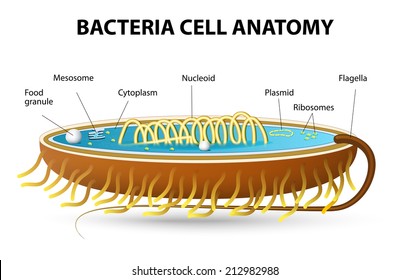
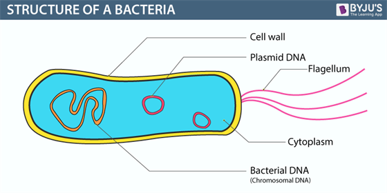
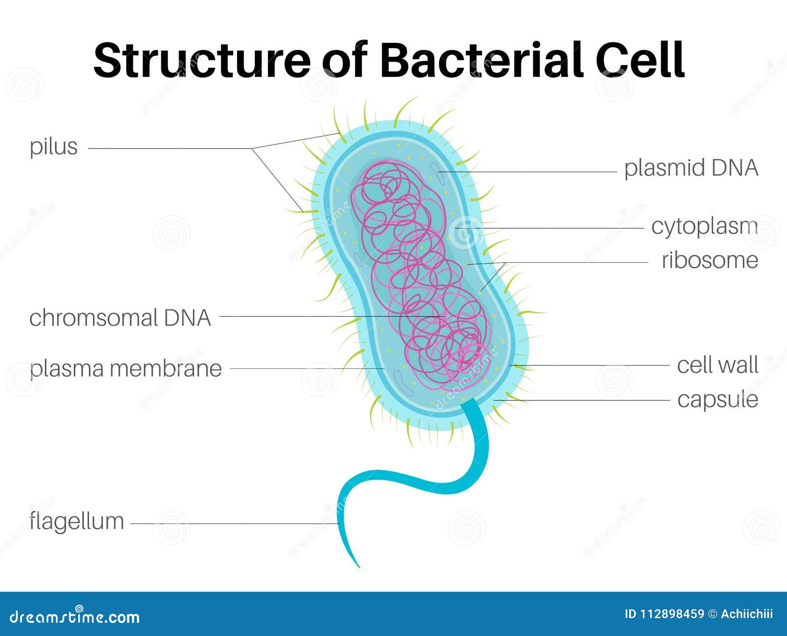
Bacteria (Prokaryotes) are simple in structure, with no recognizable organelles. They have an outer cell wall that gives them shape. Just under the rigid cell wall is the more fluid cell membrane. The cytoplasm enclosed within the cell membrane does not exhibit much structure when viewed by electron microscopy. How to draw bacteria...easy outline diagramBUY THE MATERIAL I USED 1. APSARA DARK PENCIL .....https://amzn.to/2XtrpyR2 CAMLIN PENCIL COLOURS....https://amzn.... The different parts of a generalized bacteria cell have been-shown in Figure 2.3 and have been described as follows: 1. Flagella:. Bacterial flagella are thin filamentous hair-like helical appendages that protrude through the cell wall and are responsible for the motility of bacteria.
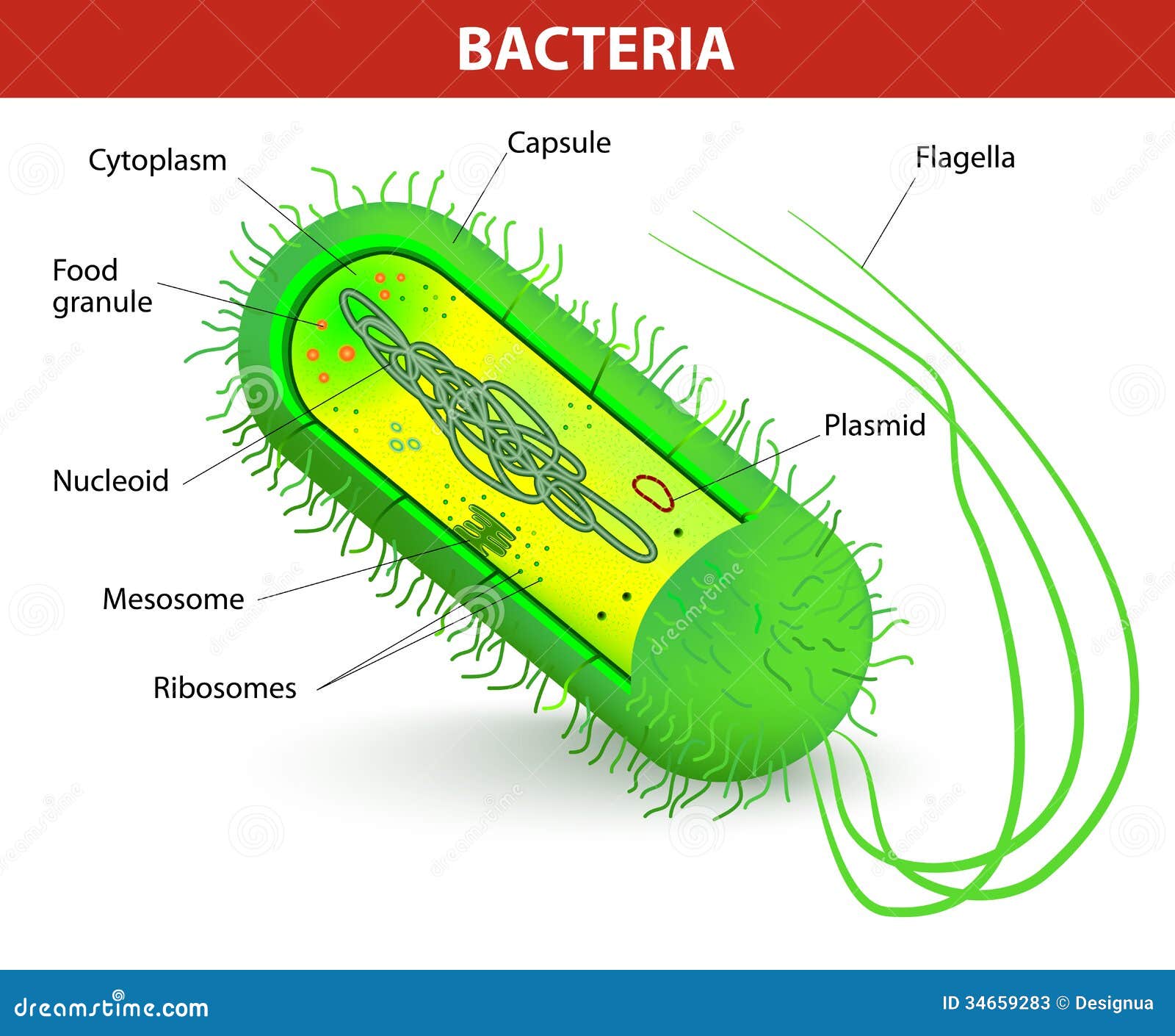
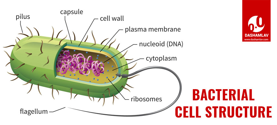
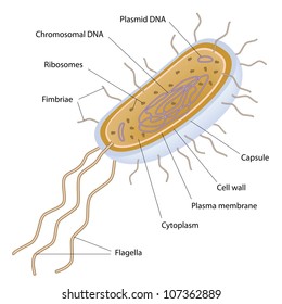
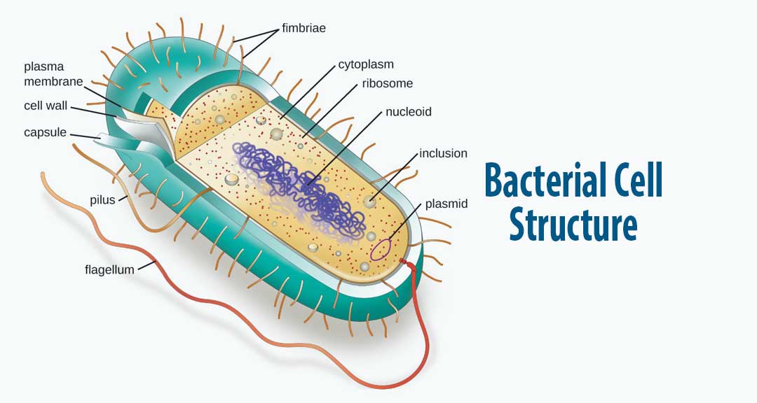
Diagram of a bacterium. There are many different types of bacteria. One way of classifying them is by shape. There are three basic shapes. 1- Spherical shape- Bacteria shaped like a ball are called cocci, and a single bacterium is a coccus. Examples include the streptococcus group, responsible for "strep throat.". 2-Rod- shape- These are known as bacilli (singular ... Structure and Function of a Typical Bacterial Cell with Diagram. Bacteria are unicellular. Their structure is a very simple type. Bacteria are prokaryotes because they do not have a well-formed nucleus. A typical bacterial cell is structurally very similar to a plant cell. The cell structure of a bacterial cell consists of a complex membrane ... English: A simple diagram of a bacterium, labelled in English. It shows the cytoplasm, nucleoid, cell membrane, cell wall, mitochondria (which bacteria lack), plasmids, flagella, and cell capsule. The source code of this SVG is valid. This diagram was created with an unknown SVG tool. This SVG diagram uses embedded text that can be easily ... Consider the diagram of the basic structure of a bacterium. A diagram of a bacterium is labeled. Part A is the cell membrane, part B is the flagellum, part C is the nucleoid, and part D is the pilus. Which of the labeled structures in the image allows bacteria to exchange genetic material and thus evolve to be better pathogens?
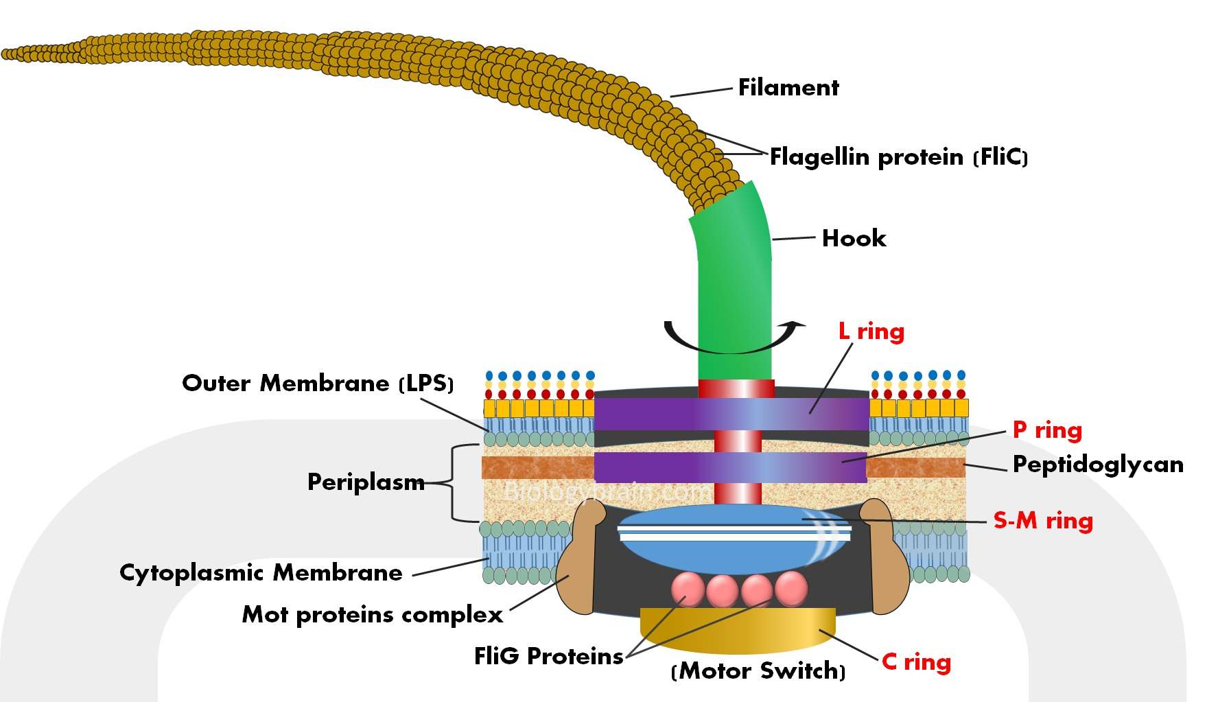
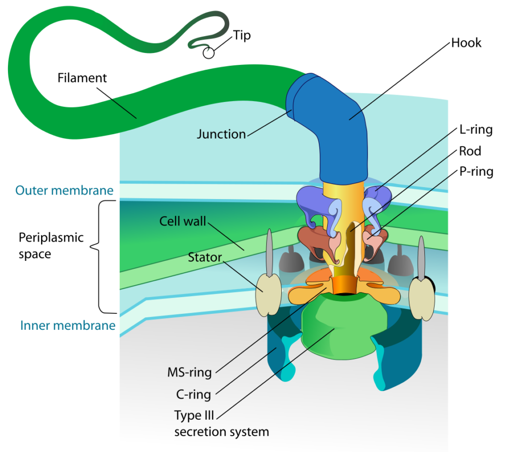
a homogeneous, generally clear jelly-like material that fills the cell. Chromosome. a very long, continuous piece of DNA, which contains genes, regulatory elements and other intervening nucleotide sequences. Ribosome. an organelle composed of rRNA and ribosomal proteins. Plasmid. Media in category "Bacteria diagrams" The following 156 files are in this category, out of 156 total. A-cartoon-of-a-bacterial-cell-intentionally-shown-uncrowded-to-highlight-the-key-living-processes-metabolism-green-cytop.jpg 358 × 281; 38 KB Sep 30, 2021 · 2,405 bacteria cell diagram stock photos, vectors, and illustrations are available royalty-free. See bacteria cell diagram stock video clips. of 25. bacteria earth prokaryotic cell bacteria diagram planet sizes bacterial cell life cycle of virus ribosomes in prokaryotic cells structure of bacterial cell flagella bacterial structure. Figure: Well Labelled Diagram of flagella of bacteria. The typical flagellum of gram-negative bacteria such as E. coli is a composite of three parts. 1. The basal body of the flagella. The basal body is a major component of the flagella of bacteria. It is a rod-shaped structure and a complex of different rings embedded in the cell membrane.
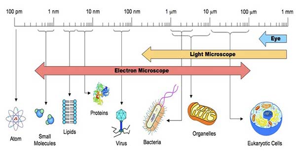
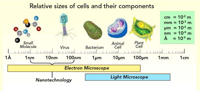
The bacterial cell diagram above shows the structures of bacterium anatomy.Bacteria are found practically everywhere on Earth and live in some of the most unusual and seemingly inhospitable places. Their size ranges between 0.2 µm and 700 µm in diameter, with the normal range of about 1-5µm in diameter. Flagellum base diagram-en.svg. English: A Gram-negative bacterial flagellum. A flagellum (plural: flagella) is a long, slender projection from the cell body, whose function is to propel a unicellular or small multicellular organism. The depicted type of flagellum is found in bacteria such as E. coli and Salmonella, and rotates like a propeller ... In this article we will discuss about the structure of bacteria. This will also help you to draw the structure and diagram of bacteria. 1. A bacterial cell remains surrounded by an outer layer or cell envelope, which consists of two components – a rigid cell wall and beneath it a cytoplasmic membrane or plasma membrane. biology questions and answers. Label The Following Diagram Of A Bacterium. Ribosome Nucleoid Fimbriae Plasma Membrane Capsule ... Question: Label The Following Diagram Of A Bacterium. Ribosome Nucleoid Fimbriae Plasma Membrane Capsule Cytoplasm Conjugation Pilus Flagellum Cell Wall.
If you start with a single bacterium capable of dividing every 30 minutes, how many bacteria would you have after just 4 hours? ... 11. In Model 2 , diagram B shows that the population size fluctuates around the carrying capacity. Considering what you know about interactions in the environment, discuss with you group some ...
1. Cover different classification schemes for grouping bacteria, especially the use of the Gram stain 2. Describe the different types of bacteria 3. Discuss bacterial structure and the function of the different bacterial components 4. Discuss the distinguishing characteristics of Gram positive and Gram negative bacteria.
This diagram depicts the numerous shapes of bacteria. Bacterial morphology diagram. Types of Bacteria. The cell wall also makes Gram staining possible. Gram staining is a method of staining bacteria involving crystal violet dye, iodine, and the counterstain safranin. Many bacteria can be classified into one of two types: gram-positive, which ...
The bacterium, despite its simplicity, contains a well-developed cell structure which is responsible for some of its unique biological structures and pathogenicity. Many structural features are unique to bacteria and are not found among archaea or eukaryotes.Because of the simplicity of bacteria relative to larger organisms and the ease with which they can be manipulated experimentally, the ...
Bacteria (sing. bacterium) are unicellular prokaryotic microorganisms which divide by binary fission. They do not possess nuclear membrane and the nucleus consists of a single chromosome of circular double-stranded DNA helix (Fig. 1.1). These are long filamentous, cytoplasmic appendages, 12-30 μm in length, protruding through the cell wall ...
With the help of a labelled diagram describe the structure of a bacterial cell. Hint: Bacteria are a type of biological cell. They constitute a large domain of prokaryotic microorganisms. The length of the bacteria is a few micrometers. Bacteria have several shapes, ranging from spheres, rod, cocci and spirals.
May 17, 2021 · Today, bacteria are considered as one of the oldest forms of life on earth. Even though most bacteria make us ill, they have a long-term, mutual relationship with humans and are very much important for our survival. But before we elaborate on its uses, let us know the structure of bacteria, its classification, and the bacteria diagram in detail.
Bacteria diagram also shows Periplasmic space, which is a cellular compartment found only in bacteria that have an outer membrane and a plasma membrane. A plasma membrane that is responsible for transporting factors such as ions, nutrients or waste across the membrane and it's a lipid bilayer. Appendages are also shown, they are basically hair ...
STRUCTURE and FUNCTION. E. coli are rod-shaped bacterium that has an outer membrane consisting of lipopolysaccharides, inner cytoplasmic membrane, peptidoglycan layer, and an inner, cytoplasmic membrane. Cell wall- this structure is made of a thick layer of protein and sugar that prevents the cell from bursting.
Bacteria that create their own energy, fueled by light or through chemical reactions, are autotrophs. Capsule - Some species of bacteria have a third protective covering, a capsule made up of polysaccharides (complex carbohydrates). Capsules play a number of roles, but the most important are to keep the bacterium from drying out and to protect ...
A diagram of a bacterium is labeled. Part A is the cell membrane, part B is the flagellum, part C is the nucleoid, and part D is the pilus. Which of the labeled structures in the image allows bacteria to exchange genetic material and thus evolve to be better pathogens?
The different parts of a generalized bacteria cell have been-shown in Figure 2.3 and have been described as follows: 1. Flagella:. Bacterial flagella are thin filamentous hair-like helical appendages that protrude through the cell wall and are responsible for the motility of bacteria.
How to draw bacteria...easy outline diagramBUY THE MATERIAL I USED 1. APSARA DARK PENCIL .....https://amzn.to/2XtrpyR2 CAMLIN PENCIL COLOURS....https://amzn....
Bacteria (Prokaryotes) are simple in structure, with no recognizable organelles. They have an outer cell wall that gives them shape. Just under the rigid cell wall is the more fluid cell membrane. The cytoplasm enclosed within the cell membrane does not exhibit much structure when viewed by electron microscopy.

Bacteria Cell Diagram Poster Wall Art School Fun Baby Shower Present Learning Gift Kids Amazon Co Uk Home Kitchen

I Identify The Following Bacteria Based On Its Shape Ii Draw And Label The Following Parts Of The Bacteria A Flagella B Pili C Cell Wall Cytoplasm Cell Membrane
Bacteria Cell Diagram Stock Illustrations 1 061 Bacteria Cell Diagram Stock Illustrations Vectors Clipart Dreamstime















Comments
Post a Comment