42 where in the diagram is the distal epiphysis
Distal Epiphysis Function Drivers! find and download drivers laptops, computer, printer The distal epiphysis is the rounded part of the bone found at the end of the diaphysis that is pointing Drivers. Details: Download scientific diagram Diagram of supportive tendons and ligaments of the equine foot... [Radiographic parameters of the distal epiphysis of the radius in the healthy and fractured wrist]. Arch Putti Chir Organi Mov.
Epiphyseal Injuries (Salter-Harris Classification). The epiphysis is a common site of injury in the growing skeleton. If the epiphysis is resected or lost secondary to tumor or trauma, respectively, growth potential can be restored in a child with skeletal immaturity through vascularized epiphyseal...

Where in the diagram is the distal epiphysis
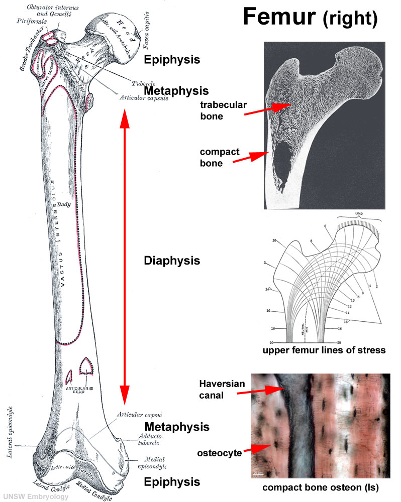
was designed to treat the fractures of distal femur meta-epiphysis with particular emphasis on supracondylar fractures. The pathological fractures can be also treated with CHARFIX2 condylar nail. Its additional advantage is the possibility of supplying diaphysis femur fractures with previously... Slipped upper femoral epiphysis is more common in boys than girls and more common in Afro-Caribbeans than Caucasians. In the pre-slip phase, there is a widening of the growth plate with irregularity and blurring of the physeal edges and demineralization of the metaphysis. The distal epiphysis is located at the end of the long bone that is farther away from the center of the body. How long does it take for an epiphysis fracture Fractures in the proximal tibia (top of the shin bone) or distal femur (bottom of the thighbone) are the most common epiphyseal plate injuries that...
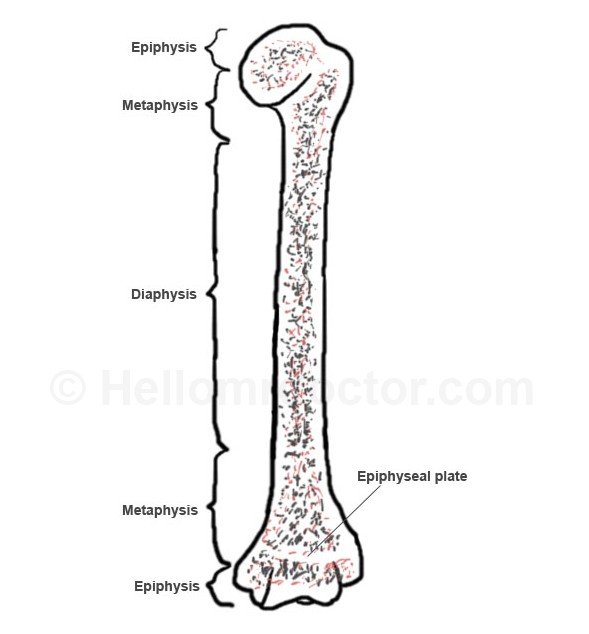
Where in the diagram is the distal epiphysis. Epiphysis Definition It is a vital growth area near the end of a long bone, which later fuses with the main bone through ossification. It is often mistaken for epiphysis cerebri, a small endocrine gland in the brain. Its plural is epiphyses. The most notable part is that the Epiphysis has red bone marrow in... Distal epiphysis Proximal epiphysis Diaphysis Metaphysis Reset. [Numbered in a sequence from top to bottom of the long bone] 1. Proximal epiphysis (put it in the top right blank box) - It is the topmost part of the long bone given in the picture. The epiphysis is the round end of the long bone. It is further categorized as the proximal In order to facilitate this function, the proximal and the distal epiphysis are covered with layers of articular cartilage. The epiphyseal line/plate in the metaphysis separates the diaphysis from the epiphysis. My house has no rooms that are all interior walls. My house is wood with a crawl space. I live in Northern Alabama. Is there any preferred room to stay in that would be best? My closets have hanging doors and wouldn't do a thing to protect. House built in 77. ​ [https://i.imgur.com/TLhRkGa.png](https://i.imgur.com/TLhRkGa.png) ​ Any suggestions?
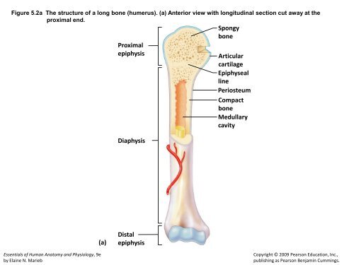
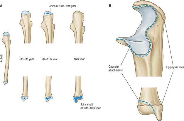
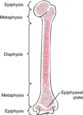
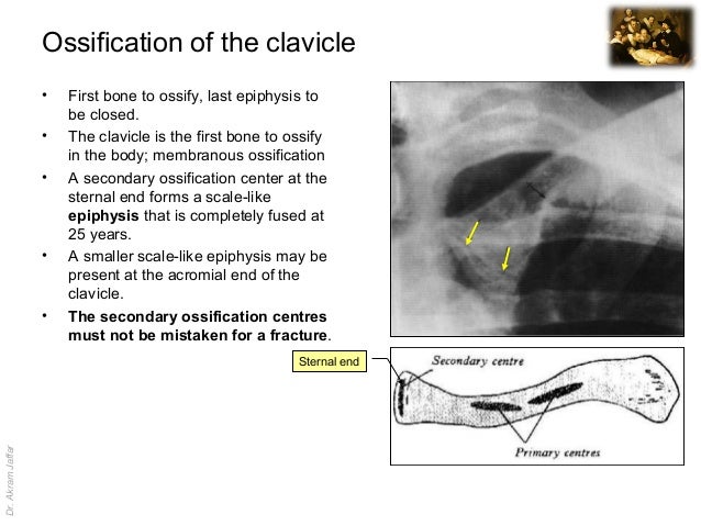
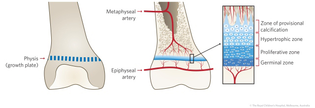
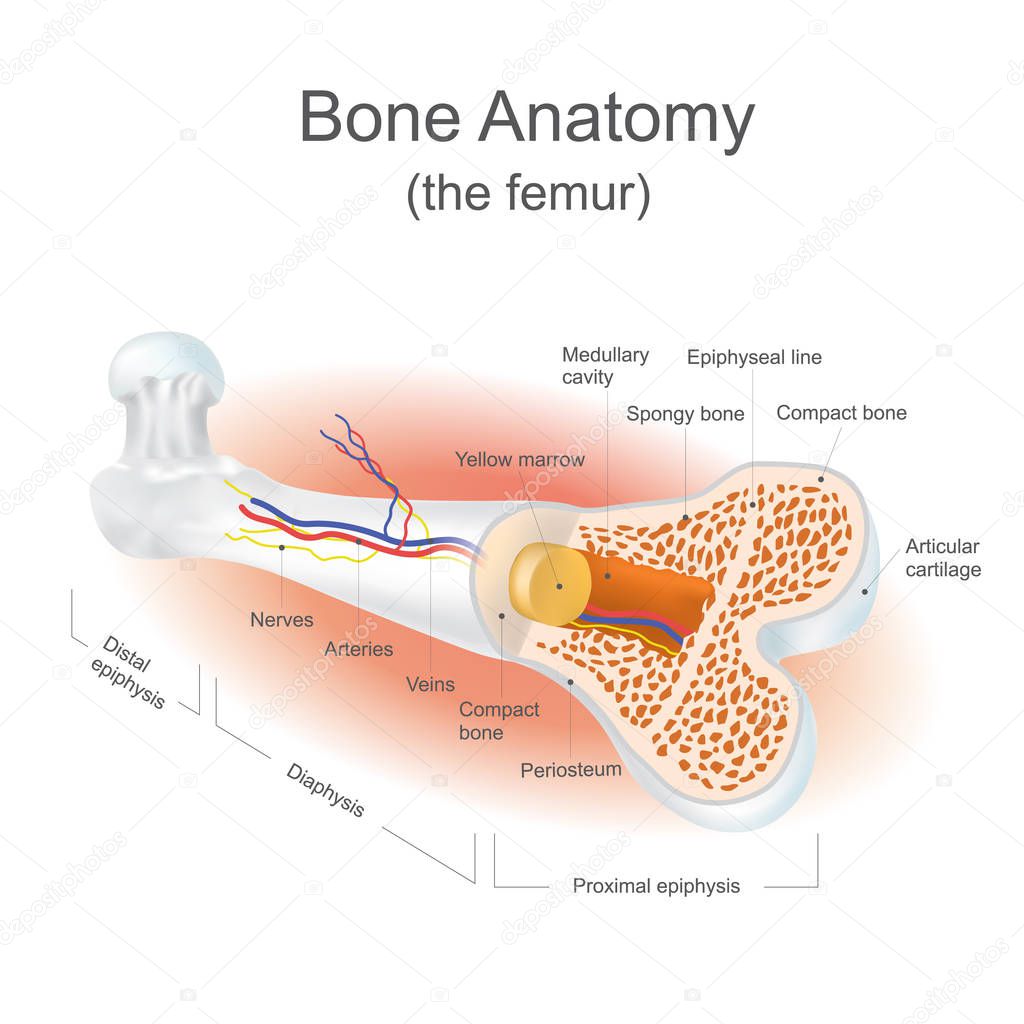
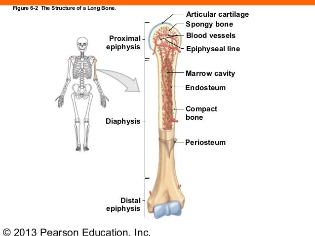
The epiphysis is the rounded end of a long bone, at its joint with adjacent bone(s). Between the epiphysis and diaphysis (the long midsection of the long bone) lies the metaphysis, including the epiphyseal plate (growth plate). A Hasse diagram is a graphical representation of the relation of elements of a partially ordered set (poset) with an implied upward orientation. If p The diagram shows development of the epiphyseal nucleus in the proximal end of the femur. Figure 2. Arterial supply of the proximal femur. The capital femoral epiphysis and physis are supplied by It is most common during the initial 2 years after fracture, especially with fractures of the distal third of... Here's what I'm trying to accomplish: http://imgur.com/a/0didk The concept of a car's scissor jack would be perfect, but this is going in a very small space, so the whole assembly has to be no wider than 0.75". Is there some small piece of hardware that accomplishes the same thing? The closest thing I've found would be an aircraft expansion bolt, but the geometry of that is a bit different and it's not robust enough to solidly control the space between two surfaces. Thanks for your help! Epiphysis is the rounded end of a long bone. Its main function is to form joints with adjacent bones. Metaphysis contains the growth plate of the epiphysis known as the epiphyseal plate. Two epiphyses occur at the proximal and distal end of a long bone while single diaphysis occurs per long... Distal tibial physeal fractures are classified by the Salter-Harris classification. They can also be Tillaux fracture (Figure 2) - a Salter-Harris type III fracture involving avulsion of the anterolateral corner of the distal tibial epiphysis (the last The distal tibia is the third most common physis to be injured. epiphysis vs diaphysis | Images are from http://www ... In the diagram, where is the epiphyseal vein? Difficulty: mediumFeedback: 6.451. In the diagram, where is the nutrient artery?a.Db.Ec.Fd.Ge.HAns: E. Difficulty: mediumFeedback: 6.4Essay52. Briefly describe what is happening in each of the stages above. Tumor limited to the distal ulnar epiphysis, enlarged ... Typical epiphyseal fracture occurs in the transformation zone and separates the growth plate, leaving proliferating cells attached to the epiphysis. Complications resulting from Salter-Harris type II fractures are uncommon, except in the distal femoral epiphysis where they occur in 43-70% of all... Distal Anatomy Distal epiphyses in anterior, distal, and posterior views. In the early nineteenth century, creationists' approaches (Cuvier-Paley) helped to install the idea of a form-function binomial that gained scientific status in the second half of that century when it was contextualized within the framework of evolution... Forearm | Basicmedical Key b) distal epiphysis. c) nutrient foramen. Where in the diagram can you find red bone marrow in an adult? Which of the labeled structures in the diagram are fragments of older osteons that have been partially destroyed during bone rebuilding or growth? Closeup of skeleton foot model Where there is involvement of the whole epiphysis, autologous epiphyseal transfer may achieve the dual goals of restoring joint function and growth potential. It develops in the bone bud, secondary to the primary ossification centers (metaphysis) and is responsible for longitudinal and circumferential... Ankle Fractures and Dislocations Injuries | Bone and Spine The newest Pic2Mag's Field Calculator version has went from Beta Version 1.0 to Free Version 1.01. Mainly polished the program up some and added a few more features. The Field Calculator Version 1.01 has better load balancing between processor threads, and now supports monopoles and extended length compass needles. http://www.pic2mag.com/Pic2Mag_FieldCalc_v101.pdf You can find the program download link in the manual, or on the Pic2Mag website. Please feel free to share, distribute, and ... Epiphysis - Definition, Location, Function and Pictures bone. Proximal. epiphysis Articular. cartilage. Epiphyseal. Diaphysis. Distal. epiphysis. Copyright © 2009 Pearson Education, Inc., publishing as Pearson Benjamin Cummings. A: Anteroposterior radiograph-subtle widening of the ... The distal epiphysis is made up of spongy bone, which is bone with tiny holes similar to lattices. These holes are filled with connective tissue and bone Below it lies the physis, the area where growth occurs. Distal to that is the metaphysis, a flared region of bone, and below that lies the narrower... Imaging anatomy fracture of the clavicle The characteristic pattern of fusion of distal tibial epiphysis explains the special configuration of the fragments in the triplane fracture and the "juvenile" fracture of Tillaux in adolescents. Eight patients, 13 to 15 years of age, with distal tibial epiphyseal fractures were treated in the last 12 years. Salter-Harris fracture classifications. Drawings show the ... Where are the epiphysis and diaphysis located? Anatomical terminology. - distal epiphysis includes entire articular surface of distal end of femur & serves as origin of part of gastrocnemius muscle; - epiphyseal center of distal aspect of femur is present at birth in newborns, & it expands... Osseous Tissue flashcards | Quizlet Distal Epiphysis. Quizlet is the easiest way to study, practise and master what you're learning. Create your own flashcards or choose from millions created by other In which region of the diagram would you find the medullary cavity? C. Where in the diagram can you find red bone marrow in an adult? 3D scan of the proximal epiphysis of a right tibia ... groove between tubercles Distal Epiphysis of the Humerus: - 3 depressions that are periodically occupied by some of the forearm bones depending on Appendicular. -To the left side of the diagram. -It is the remaining elements of the skeletal system. - Consists of : Bones in the appendages: Bones... This illustration depicts an anterior view of the right ... Epiphysis Definition - Epiphysis is the rounded end of a long bone, its primary function is to connect adjacent bones to form joints. There is another part of the long bone between the epiphysis and the diaphysis, which we call metaphysics. The epiphyseal plate, or growth plate of the epiphysis, is... A8-22, right distal epiphysis of humerus in cranial (1a ... Can someone explain that to me a little better? What’s the *distal* ureter? I had an x-ray a few days ago in preparation for a ureteroscopy this friday for the 1 cm stone. I’ve been passing this stone for nearly 10 months but i’d really like to put any procedure off as long as possible. I plan on asking my doctor about this it’s just that Im unable to speak to him until tomorrow, I saw my results on my patient portal and I’m really curious. I’m in denial and think i’ll pass it 🤣 US images. (A) The distal epiphysis of the MTHs is marked ... So the PS pump is going out on my 2006 acura rsx type s. I tried replacing the o-ring on the inlet, and it still sounds horrible and sounds like it's going out, so I need to replace the pump. A new pump is $650, so instead I bought a used one from the junk yard for $70 and I bought a set of new o-rings, seals, and bearings from the dealership for $20. I watched a lot of videos on rebuilding pumps and set to work with a diagram of the disassembled pump. My only issue is, after I pulled the pump s... Fracture Education : Physeal (growth plate) injuries The distal epiphysis is the growth plate of the long bone farthest from the body. Add your answer: Earn +20 pts. Q: Where is the distal epiphysis? File:Bone-femur.jpg - Embryology The distal epiphysis is made up of spongy bone, which is bone with tiny holes similar to lattices. These holes are filled with connective tissue and bone marrow. Details: The epiphysis is the area of the long bone where bone growth takes place. Long bones actually grow from the inside out. Bone terminology diagram | Image | Radiopaedia.org Where in the diagram is the distal epiphysis? 50. Where in the diagram is the only place that does not have a periosteum? Use the following to answer questions 51-54 51. Long Bone Anatomy: Structure and Parts of Long Bones Where is the parts diagram for the power steering fluid reservoir? https://estore.honda.com/honda/parts/view-honda-parts-catalog.asp?m=2005-cr-v-5-lx-4wd-5at&dl= hello yellow on a small wall A periosteum b distal epiphysis c nutrient. C where in the diagram can you find red bone marrow in an adult. It contains numerous chondrocytes scattered in a The distal epiphysis is described as the rounded end of the bone located at the end part of the diaphysis which is located away from the... | Distal femoral metaphyseal and epiphyseal trabecular ... The distal epiphysis is located at the end of the long bone that is farther away from the center of the body. How long does it take for an epiphysis fracture Fractures in the proximal tibia (top of the shin bone) or distal femur (bottom of the thighbone) are the most common epiphyseal plate injuries that... Diagram showing the growth of epiphysis of contralateral ... Slipped upper femoral epiphysis is more common in boys than girls and more common in Afro-Caribbeans than Caucasians. In the pre-slip phase, there is a widening of the growth plate with irregularity and blurring of the physeal edges and demineralization of the metaphysis. place to be was designed to treat the fractures of distal femur meta-epiphysis with particular emphasis on supracondylar fractures. The pathological fractures can be also treated with CHARFIX2 condylar nail. Its additional advantage is the possibility of supplying diaphysis femur fractures with previously... 28 Where In The Diagram Is The Distal Epiphysis - Wiring ... Fragments of the distal epiphyseal cartilage of the femur ... Closeup of skeleton pelvic model TRAP and CD68 staining in the distal femoral epiphysis. (A ... Solved: Label The Regions Of A Long Bone. Distal Epiphysis ... Radiograph of the distal radius and ulna showing the U5 ... Physeal Injuries and Growth Disturbances | Musculoskeletal Key 0 Typical chondroblastoma of the distal femoral epiphysis ... Closeup of skeleton hand model Eames Chair Diagram Schematics - 1951 Chair Poster Hanging on Wall Volar view of the distal radius showing, in dotted line ... Femoral (mean ± SD) trabecular bone of the distal ... Skeletal system lecture exam Flashcards | Easy Notecards Long Bone Labeled Periosteum : Bone Development And Growth ... Long Bone Diagram Epiphyseal Plate / Cartilage Bone ... Places To Be , Hamburg Metaphysis - Anatomy, Pictures, Clinical Significance and ... Long Bone Diagram Epiphysis - Diagram Media 163 ch 06_lecture_presentation


Comments
Post a Comment