43 vocal folds diagram
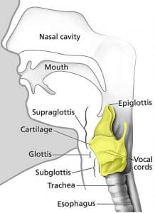
Vocal cords: structure and function | Kenhub The vocal folds are wedged-shaped structures consisting of the vocal ligament, the vocalis muscle and a mucous membrane covering. They span the laryngeal cavity in an anterior-posterior direction and have a long free edge that protrudes into the laryngeal cavity. In a relaxed state, a small slit-like space can be found between the two folds. Larynx (Voice Box) Definition, Function, Anatomy, and Diagram Cartilage Structure of Larynx. Its internal cavity can be divided into the following parts: Supraglottis: The part above the vocal cords, containing the epiglottis [4] Glottis: The area consisting the vocal cords or folds; there are two pairs of vocal folds (mucous membrane structures) in the larynx, the false vocal folds and the true vocal folds [5].The former is covered with respiratory ...
Vocal Cord Photos and Premium High Res Pictures - Getty Images vocal cord diagram vocal cord polyp 341 Vocal Cord Premium High Res Photos Browse 341 vocal cord stock photos and images available, or search for vocal cord anatomy or vocal cord diagram to find more great stock photos and pictures. of 6 NEXT

Vocal folds diagram
Anatomy of the Vocal Folds | Structure & Function | Common ... The vocal folds are intricately layered, which is the reason for unique sound production. The outer layer is squamous, non-keratinizng epithelium. Just deep to this is the superficial layer of the lamina propria. This is a gel like layer, which allows the vocal fold to vibrate and produce sound. The deeper layers of the vocal fold are muscles ... Diagrams of the larynx and vocal folds. (a) Midsagittal ... Four views of the larynx and vocal folds are shown in Fig. 2. The first is a midsagittal sketch of the head and neck indicating the location of the larynx (Fig. 2a) and the second is a schematic... PDF Vocal Folds Diagram - stats.ijm.org vocal-folds-diagram 1/20 Downloaded from stats.ijm.org on April 6, 2022 by guest Vocal Folds Diagram Getting the books Vocal Folds Diagram now is not type of inspiring means. You could not unaccompanied going subsequent to books hoard or library or borrowing from your associates to open them. This is an no question simple
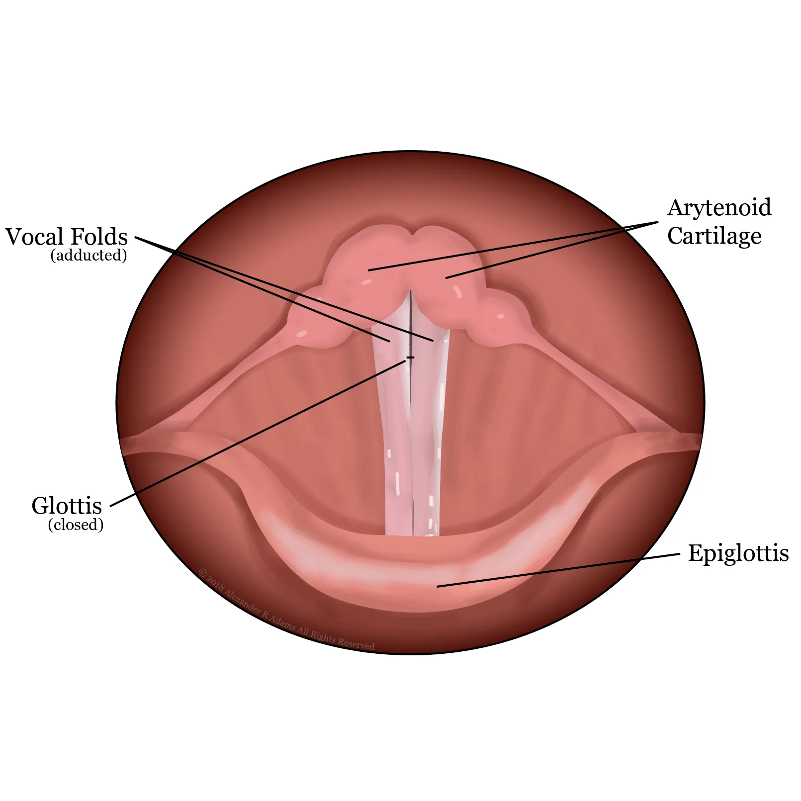
Vocal folds diagram. Label the parts exercise | Phonetics nasal cavity hard palate velum uvula oral cavity alveolar ridge dorsum blade tip teeth lips pharynx epiglottis larynx vocal folds esophagus trachea: Click on the button that matches the speech organ shown. PDF 3 Mechanisms of Voice Production - University of Arizona Figure 3.2 diagrams of the larynx and vocal folds. (a) Superior view of larynx when the vocal folds are abducted, as during respiration. (b) Superior view of larynx when the vocal folds are adducted, as during phonation. (c) division of the vocal fold into the cover and body portions (based on Hirano 1974). Understanding Voice Production 1 Column of air pressure moves upward towards vocal folds in "closed" position 2, 3 Column of air pressure opens bottom of vibrating layers of vocal folds; body of vocal folds stays in place 4, 5 Column of air pressure continues to move upward, now towards the top of vocal folds, and opens the top New vibratory cycle 6-10 The low pressure created behind the fast-moving air column ... The Voice Mechanism - THE VOICE FOUNDATION Vocal Folds. The left and right vocal folds are housed within the larynx. The vocal folds include three distinct layers that work together to promote vocal fold vibration. Covering/mucosa: Loose structure that is key to vocal fold vibration during sound production; is composed of: Epithelium; Basement membrane; Superficial lamina propria (SLP)
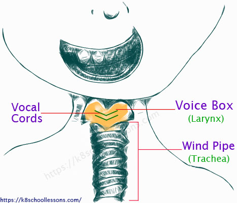
Schematics of the vocal folds. | Download Scientific Diagram Download scientific diagram | Schematics of the vocal folds. from publication: Modeling vocal fold asymmetries with coupled Van der Pol oscillators. | Models of the glottal sound source are being ... Laryngeal Ligaments and Folds - Vocal - Vestibular ... Vocal ligament - Lies at the free upper edge of the cricothryoid ligament. Vocalis muscle - Exceptionally fine muscle fibres that lie lateral to the vocal ligaments. The vocal folds are relatively avascular, and appear white in colour. The space between the vocal folds is known as the rima glottidis. Structure and Functions of the Vocal Cords Explained With ... The vocal cords, which are also referred to as the vocal folds, are situated within the larynx (voice box), which is placed on top of the trachea (windpipe). The muscles from the hyoid bone (U-shaped bone at the base of the tongue) support the larynx, enabling it to move up or down. The larynx consists of 3 paired and 3 unpaired cartilages. Vocal Folds Diagram | Quizlet Start studying Vocal Folds. Learn vocabulary, terms, and more with flashcards, games, and other study tools.
vocal folds labeling Diagram | Quizlet Vocal Folds Terms in this set (9) Vallecula ... Root of tongue ... Epiglottis ... Piriform Sinus ... Vocal fold ... Cuneiform tubercle ... trachea ... ventricular fold ... s aryepiglottic fold THIS SET IS OFTEN IN FOLDERS WITH... Larynx Labeling 9 terms veraav vocal tract labeling 7 terms veraav Larynx anterior and posterior labeling 7 terms veraav Vocal Cords Illustrations, Royalty-Free Vector Graphics ... Browse 141 vocal cords stock illustrations and vector graphics available royalty-free, or search for the vocal cords to find more great stock images and vector art. Newest results the vocal cords Larynx anatomical vector illustration diagram, educational... Vocal cords labeled anatomical and medical structure and... The larynx Anatomy, Head and Neck, Larynx Vocal Cords - StatPearls ... The vocal folds are now widely agreed upon and understood facets of voice because of modern histological techniques. The vocal fold comprises five layers (deep to superficial layers as follows): thyroarytenoid muscle, deep lamina propria, intermediate lamina propria, superficial lamina propria, and the squamous epithelium. Vocal Cords Diagram - Lan Diagrams Vocal Cords Diagram. Animals also have vocal folds, which allows them to vocalize. A vocal EQ cheat sheet to help you mix vocals like a pro. Good day! Welcome! :D: The Origins Of Language (Jason Morrison) The glottal consonant [h] is articulated in the glottis. The vocal cords (or vocal folds) are located in your larynx (voice box).
Vocal Folds Diagram | Etsy Check out our vocal folds diagram selection for the very best in unique or custom, handmade pieces from our shops.
Vocal Tract: Anatomy & Diagram | Study.com Learn about the anatomy of the vocal tract, which is the part of the body responsible for producing human speech. Examine the roles of the larynx and the pharynx, and see a diagram of the vocal tract.
Muscles of the larynx: Anatomy, function, diagram | Kenhub Vocal fold paresis The recurrent laryngeal nerve is responsible for innervating all muscles of the larynx except the cricothyroid muscle. The clinical term to describe when one or two of the recurrent laryngeal nerves are injured is vocal fold paresis (also known as recurrent laryngeal nerve paralysis or vocal fold paralysis).
Voice & Swallowing - Anatomy - OHSU The vocal folds lie in the center of this structure in a front to back alignment. When viewed from above the vocal folds appear as a "V"-shaped structure with the opening between the "V" being the entrance to the trachea (wind pipe, air tube). At the rear of the larynx on each side, each vocal fold is attached to a small arytenoid cartilage.
Vocal cords - Simple English Wikipedia, the free encyclopedia The male vocal folds are between 17 mm and 25 mm in length. The female vocal folds are between 12.5 mm and 17.5 mm long. The difference in vocal fold size between men and women means that their voices have a different pitch. Each person's voice is different and has a slightly different pitch.
diagram and vocal folds | جدني diagram and vocal folds. في هذه الصفحة سوف تجد مواضيع عن vocal cord bones وpicture of false vocal cords، بالإضافة إلى glottis anatomy illustration وlarynx with glottis closed، كذلك vocal chords anatomy، علاوة على صفحات في true vocal cords anatomy model، أيضا epiglottis glottis و epiglottis and glottis، بإلإضافة إلى ...
Vocal Cords Stock Photos, Pictures & Royalty-Free Images ... Vocal cord anatomy vector illustration diagram, educational... The vocal folds, known as vocal cords, are two folds of muscle... Human larynx anatomy The structure of the vocal cord. A teacher holds a sore throat in a school classroom. Voice... Throat Anatomy Human larynx Middle-aged Japanese woman holding her neck Intubation tube in airways.
The Vocal Folds / Vocal Cords | Vocalist There are two types of vocal folds (also known as vocal cords) which are referred to as 'true' and 'false'. The latter protect the more delicate 'true' folds and are located just above them. True vocal cords are two pieces of tissue which are located above the windpipe and stretch horizontally across the larynx.


Comments
Post a Comment